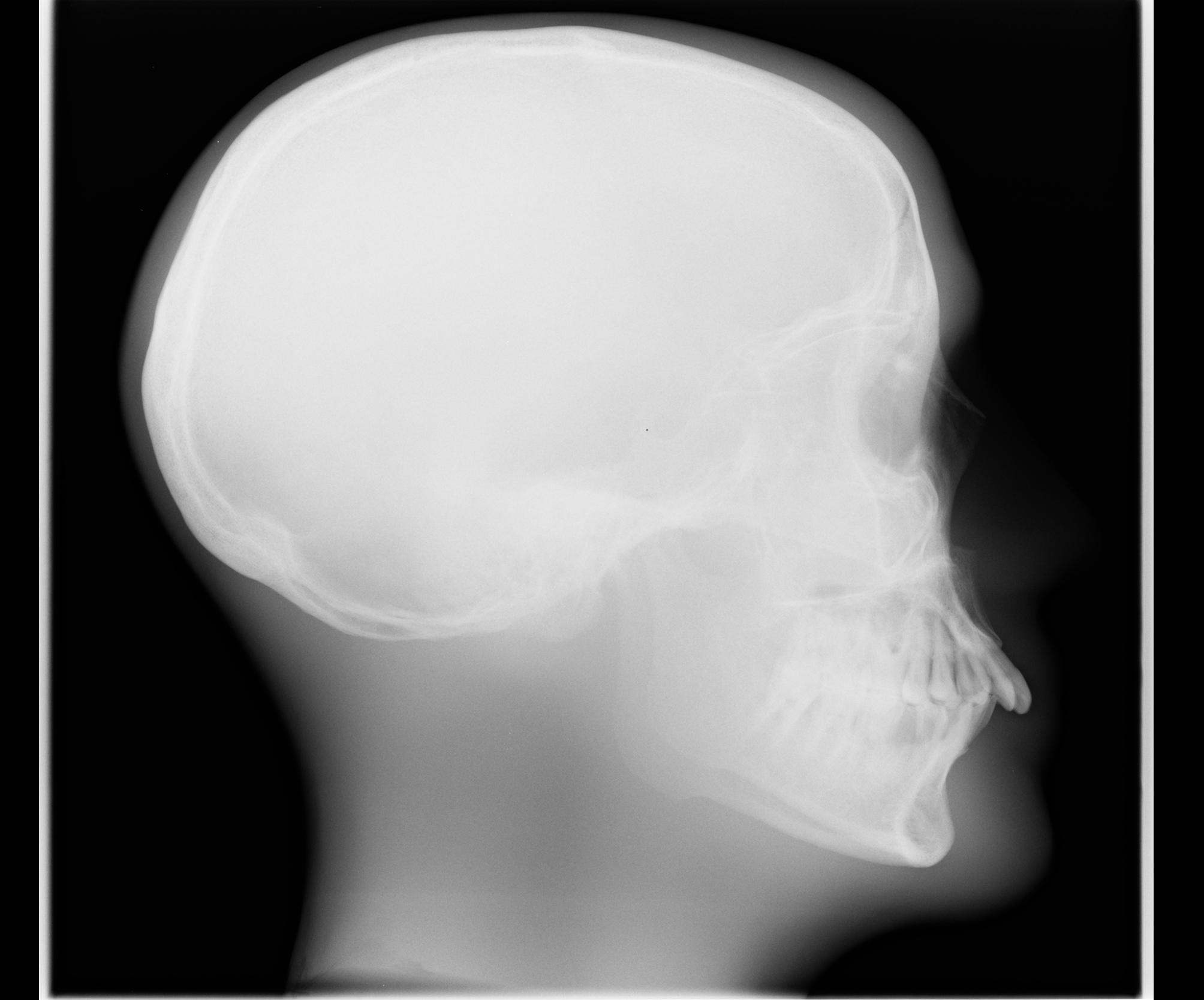
In the range of kVp less than 80 a change of two kVp for each cm change in thickness will maintain a quality image. With exception of cats and small dogs a grid or buckey must be used.
On the other hand in radiography the kVp values ranged from 55 to 65 at.

Normal kvp and low mas radiograph. Milliampere-seconds also more commonly known as mAs is a measure of radiation produced milliamperage over a set amount of time seconds via an x-ray tubeIt directly influences the radiographic density when all other factors are constant. MAs too low underexposure results in a pale film. This requires a very low kilovoltage potential kVp and corresponding relatively low milliamperage-seconds mAs.
Detector that receives greatest amount of exposure. What does kVp and mAs do. A shoulder or knee is thicker and denser than the finger and therefore more kVp is needed to adequately penetrate.
How does kVp and mAs affect image quality. Mass density of the part must be estimated. From a technical standpoint thoracic radiographic exposure should be obtained using a high peak kilo-voltage kVp 80120 kVp and low milliampere sec-ond mAs 15 mAs technique.
Compound to a low ratio grid a high grid will do which of the following. The exposure is repeated on the contralateral nasal bone for comparison purposes. Several examples would include.
By increasing or decreasing the x-ray beam current mAs which influence the amount of darkness on the image. The number of x-ray photons produced by the x-ray tube at the setting selected quantity of x-rays. A setting of between 55 and 60 kVp is typically selected.
A setting in the 65-75 kVp range is usually selected for these body structures. An increase in tube current mA results in a higher production of electrons that are inside the x-ray tube. Extremities use low kVp because they are small.
-should terminate at a max of 600 mAs at 50 kVp. Tube voltage in turn determines the quantity and quality of the photons generated. Governs the amount of X-Rays reaching the film.
Learn vocabulary terms and more with flashcards games and other study tools. In the fluoroscopy the kVp factors ranged from 45 to 55. Short scale contrast images that appear black and white is created by using a high mAs and low kVp.
Kilovoltage peak kVp is the peak potential applied to the x-ray tube which accelerates electrons from the cathode to the anode in radiography or computed tomography. KVp stands for kilovoltage peak. Image magnification in fluoroscopy helped in close up-view but.
The skin dose is now 32 mGy which is lower than the 76 mGy associated with the radiograph taken at 60 kV. Start studying Image Acquisition Radiographic Exposure Technique. It reduces pt dose.
The following variables influence the x-ray beampri-mary signal. Technical characteristics for abdominal radiography include low KVP 60-80 KVP and consequently high mAs settings. Anything that is foreign to the normal radiographic image is called what.
For facilities using film-screen this same technique can be used with a detail cassette in the upright Bucky in lieu of a computed radiography imaging plate. The film is more uniformly grey a flat film mAs. For a more penetrating beam less radiation is required at the patient entrance to achieve the required intensity at the imaging plate ie 7 mGy.
Same thickness doesnt equal same technique. With low mA values of 12 to 17. Between 80-100 kVp the change is three kVp for each cm change in thickness.
However this causes a reduction in image qualityCNR. Higher kVp and low mAs is preferred because. Energy Frequency Wavelength Number of x-ray photons produced Penetrability.
A greater increase is needed for high kVp 90 and above than for low kVp below 70. Remember that a 15 change in kVp does not produce the same effect across the entire range of kVp used in radiography. How long the exposure lasts.
Chests use high kVp low mAs they are high contrast however our technique gives us a low and long scale of contrast. Low mAs produces too light underexposure film. Tissues appear black or white with very little grey soot and whitewash Films taken with a high kV have low contrast ie.
Recommendations The 300-mA machine may have adjustable mA stations of 25 50 100 200 and 300 and two 1 and 2 mm or smaller focal spots. The hip abdomen and pelvis are even thicker and denser than the knee or shoulder. This allows you to make a variable kVp chart for the abdomen.
The image shown above was generated at 120 kV and required an exposure of only 6 mAs. In addition whenever a 15 change is made in the kVp to maintain the exposure to the IR the radiographer must adjust the mAs by a factor of 2. This technique allows for latitude long gray scale images which are impor-tant when evaluating the structures of the thorax.
The mAs used depends on the speed of the intensifying screenfilm combination. For example a kVp of 40 or 45 is used for the smallest patients which is the lowest allowable setting on diagnostic x-ray units. Constant mAs milli-ampere seconds of 10.
Low kV Coronary CTA 100 patients 85 kg Dual Source 64 CT Retrospective gating 120 kVp 330 mAs. HIPPELVIS Grid mAs CM kVp mAs CMkVp kVp AP HipPelvis Y 15 13-14 72 30 19-20 78 25-26 84 44 225 15-16 72 45 21-22 78 27-28 84 30 17-18 72 60 23-24 78 29-30 84 KNEE APOblq Knee Grid mAs CM kVp Yes 113 7-8 66 150 11-12 70 15-16 70 44 150 9-10 66 225 13-14 7017-18 Lateral Knee Decrease 4 kVp Decrease 4 kVp Decrease 4 kVp LOWER LEG APLateral Grid mAs. 12 mSv 100 kVp 330 mAs.
An increase in kVp extends and intensifies the x-ray emission spectrum such that the maximal and. 15 In the case of distal extremity exposures it could be argued that despite the kVp being lowered the. Soft tissue radiography uses low kVp and high mAs because they are low subject contrast.
KVp and low mAs. For kVp greater than 100 the change must be four kVp for every cm change. A An artifact B A blemish C A foreign body.
The power and strength of the x-ray beam quality of the x-rays. By using this technique the inherently poor abdominal contrast differences are enhanced and abdominal detail is increased. 2-Overpenetrated too much kVp or Underpenetrated too little kVp.
A Absorb more primary radiation. 8 mSv 39 decrease in radiation exposure Pflederer T et al. 1-Overexposure too much mAs or Underexposure too little mAs.
Doubling radiographic density requires doubling the mAs or increasing the kVp by 10 in the 40- to 100-kVp range or 15 in the 100-kVp or greater range. Similar to those of a finger exposure technique ie low kVp low mAs and small focal spot. Radiographic dose optimisation strategies for lowering dose are traditionally based around increasing kVp and decreasing mAs around a constant detector exposure high kVp technique.
This technique is used for imaging the abdomen and lung. If a non-grided cassette is used the kVp or penetration is lowered otherwise if the selected kVp is too low the contrast is increased resulting in a few shades of gray radiograph may appear penetrated but the mediastinum of the chest appears underexposed. Films taken with a low kV have high contrast ie.
Fixed kVp and fixed mAs B Fixed kVp and variable mAs C Fixed mAs and variable kVp. A high penetration kVp of xray is usually use demonstrate all thoracic anatomy on the radiograph. The 25 rule states that a 25 increase in mA s or mAs is required for each centimetre the patient is greater than the average2030 This is more of a guideline as it only works for radiographic situations low kVp with no grid and high kVp with grid and is an average of values that deviate from this by 25 depending on the kVp3235 Manufacturers.

Scatter And Kv Radiology Suny Upstate Medical University

Tidak ada komentar