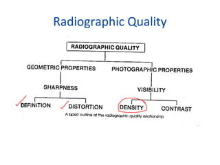
Rādē-ō-grafik densi-tē The amount of blackening on an x-ray film produced by the interaction of silver halide crystals with developing agents. Many densities or shades of gray.
The goal in dental radiology is to use techniques that require the least amount of radiation exposure to produce imageswith the right amount of density and contrast.

Radiographic density definition dentistry. They provide information about disease though its radiographic features. In addition they are helpful in treatment planning and patient management. D log I0It Density Types too white Radiographic density too white.
Non-uniform image density. Computers A measure of the number of bits that can be stored in a given amount of physical space on a storage medium. Acrylic typically does not possess any opaquers and therefore is usually totally radiolucent.
A radiographic image is formed by a controlled burst of X-ray radiation which penetrates oral structures at different levels depending on varying anatomical densities before striking the. Two or more radiographic areas on the image that exhibit different densities for same structures. Line pairs testing devices.
1 RADIOGRAPHIC DENSITY The overall degree of darkening of an exposed film is referred to as radiographic density. An artifact is a structure or an appearance that is not normally present on the radiograph and is produced by artificial means. Medical Dictionary for the Health Professions and Nursing Farlex 2012.
Radiographic examination is still left much. Radiographic interpretation is the process in which information is gleaned from radiographs. Radiographic errors may be due to technical errors errors related to the technique of taking the radiograph or processing errors related to all aspects of processing.
Measured as the optical density of an area of an x- ray film where OPTICAL DENSITY Log 10 Io Io - intensity of incident light from view box It - intensity of light transmitted through the film. When the radiographic density is optimum the image is both dark enough and light enough for. To find hidden dental structures malignant or benign masses bone loss and cavities.
The mass per unit volume of a substance under specified conditions of pressure and temperature. Contrast is the difference in lightness and darkness between areas on a radiograph. A readable text part on the radiographic image due to the density differences.
Dental radiographs are commonly called X-rays. 1 RADIOGRAPHIC ERRORS AND ARTIFACTSNeill Serman. The classic definition can be explained with the equation Radiographic contrast is equal to the product of the Subject times the film contrast.
This is the darkness or the black areas seen on the radiograph the soft tissue or the lack of hard tissue can be identified by Black regions on the radiograph. Partial ambient light exposure of the plate generally because of the overlapped unsheathed plates and fading on the exposed area. Dentists use radiographs for many reasons.
We will discuss in a later unit how contrast is. The intensity of light incident on the film I0 and the intensity of light transmitted through the film It. Radiography is a main diagnostic tool for detecting dental and maxillofacial lesions1 2 3Radiologic images have two dimension of three dimensional reality hence the images of different anatomical structures are superimposed on each other and thus make it difficult to detect the lesions 2 4 5.
The same sound is more intense if you hear it in a smaller area. Density is primarily determined by mA - Board question. Dental radiology includes the periapical film PAX to visualize periapical pathology bitewing films to identify occlusal and interpromimal dental caries occlusal films most commonly to identify submandibular sialolithiasis the panorex panoramic radiograph or orthopantomogram is a two-dimensional view of the bones and dentition of the upper and lower dental arches.
-overall sharpness of image. The intensity Intensity - The amount of energy a sound has over an area. What is determined by the thickness density composition atomic Definition.
Radiographic density is the logarithm of two measurements. Black areas on a developed radiograph are produced by deposits of metallic silver in the film emulsion that result from exposure to x-rays and their subsequent processing. Radiographic or Optical Density The overall blackness of the image is referred to as the radiographic density or optical density OD.
An image produced by radiation usually by x-rays and recorded on a radiosensitive surface such as photographic film or by photographing a fluoroscopic image. High suject densty low radiographic density. X-ray pictures can show cavities cancerous or benign masses hidden dental structures such as wisdom teeth and bone loss that cannot be seen during a visual.
At first glance there is little radiographic evidence of significant or periradicular change. This ratio is the inverse of transmittance. In routine radiography the useful range of density varies from 03 to 20 density.
Metals like amalgam and gold appear with a standard amount of radiopacity. Different shades of density in a radiograph. The radiographic density of composites varies the most because the manufacturers may place a different amount of barium salts or other opaque filling materials.
You can easily see fingers behind the film. The quantity of something per unit measure especially per unit length area or volume. Is the logarithm of two measurements.
Density is the overall darkness blackness of an image. A A well-angulated periapical radiograph of the maxillary right first molar taken during a diagnostic appointment for endodontic evaluation of the maxillary right quadrant. -ability to observe the fine structural lines.
How to evaluate radiographic detail. 14 line pairs per mm. The density below 03 is due to the density produced by the base and by some fog on the film base plus fog.
This equation applies to film screen radiography but not necessarily to digital radiography. Radiographs are an adjunct to the clinical examination and form part of the diagnostic process. A radiograph that has many black areas and is dark when viewed has high density.
Radiographic density Density - The mass of a substance per unit volume. Dental radiographs commonly referred to as X-ray films or informally X-rays are pictures of the teeth bones and surrounding soft tissues to screen for and help identify problems with the teeth mouth and jaw. Thickness of soft tissue or bone in a patient.
Radiographic density is defined as the degree of blackness or darkness on a radiograph. Radiographic Density - the overall amount degree of darkening on a radiograph. Radiographic contrast radiographic density radiographic definition radiographic anatomy radiograph rādē-ō-grăf n.

Radiographic Quality Chapter 5 Radiographic Quality O Refers

Tidak ada komentar