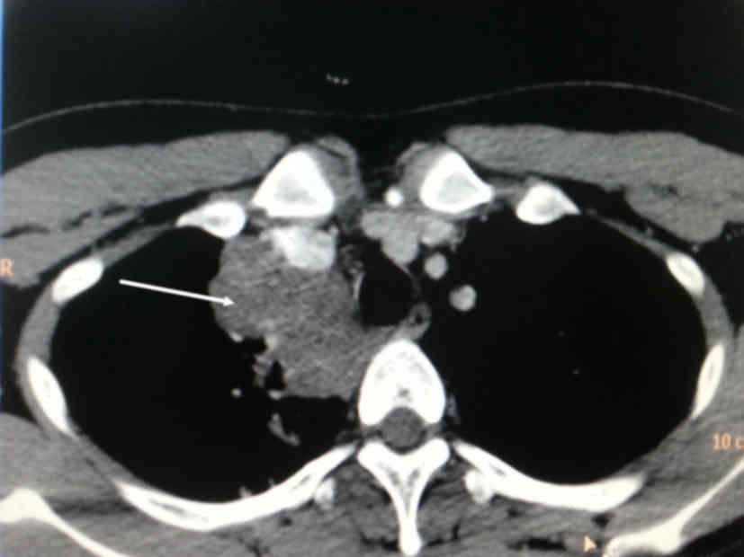
Mediastinal presentation is extremely rare. Pulmonary infarction mimicking a lung mas s.

Adult Community Acquired Pneumonia With Unusually Enlarged Mediastinal Lymph Nodes A Case Report
Tion was cr itical.
Pneumonia mimicking mediastinal mas. Lesions in the middle mediastinum may compress blood vessels or airways causing the superior vena cava syndrome Regional spread Lung carcinoma is the leading cause of cancer-related death worldwide. It is mostly found between the left lower lobe of the lung and the diaphragm. The layer that covers the lungs lies in close contact with.
Patient With Slow-Growing Mediastinal Mass Presents With Chest Pain and Dyspnea. An ascending aortic aneurysm 5 cm may mimic an anterior mediastinal mass. This led to the delay in arriving at the correct diagnosis.
A 52-year-old white woman presented with severe pain over the right upper abdomen and nonpleuritic right-sided lower chest-wall pain. Pulmonary artery dissection mimicking mediastinal mass. A number of mediastinal reflections are visible at conventional radiography that represent points of contact between the mediastinum and adjacent lung.
Cite this article. It can be visualized by echocardiography computed tomography or magnetic. Nosocomial pneumonia and sepsis Pseudomonas aeruginosa ev olved and.
Her chest CT scan revealed a. Symptoms and signs also depend on location. Interestingly mass location tends to predict malignancy.
Often an incidental finding in a patient without symptoms an ascending aortic aneurysm can cause symptoms due to compression or rupture. Thymomas lymphomas and germ cell tumors are the most frequently diagnosed tumors of the anterior mediastinum with a relative incidence of 30 20 and 18 respectively 2. Tissue sampling is necessary to diagnose cavitary lesions and to choose the right treatment.
Case presentation A 20-year-old white man a student presented to our clinic with a history of exertional breathlessness non-productive cough and fatigue over the previous 2 months. Pan African Medical Journal. We describe two identical cases of extrapulmonary sequestration mimicking mediastinal cystic mass in two boys.
Thymomas are the most common neoplasm of the anterior mediastinum with an incidence of 015 cases per 100000 3. Her pain had progressively gotten more frequent and severe over the last 5 months. Pulmonary hamartoma mimicking a mediastinal cyst-like lesion in a heavy smoker Andrea Borghesi Andrea Tironi Mauro Roberto Benvenuti Francesco Bertagna Maria Cristina De Leonardis Stefania Pezzotti Giordano Bozzola Respiratory Medicine Case Reports 2018 25.
We present the case of a young man with a primary mediastinal immature teratoma that caused compression of the main pulmonary artery mimicking pulmonary stenosis. Primary pulmonary meningioma PPM is an extremely rare benign tumor. Predisposing factors include atherosclerosis trauma vasculitis collagen-related disorders and infection.
- 2581-4214 Online ISSN No- 2581-4222 Article DOI No- 1018231 IP Indian Journal of Immunology and Respiratory Medicine-IP Indian J Immunol Respir Med. Air can enter the mediastinum. The pus-like material aspirated from these mediastinal nodes was probably inflammatory eudate similar to that.
1Different lung diseases have radiologic signs and symptoms simulating lung cancer making diagnosis difficult. A CT chest with contrast is often necessary to better identify the anatomy including the compartment affected. Percutaneous and surgical biopsies while suggesting a potential epithelial malignancy were non-conclusive.
This was followed by chest computed tomography CT that showed bilateral diffuse mediastinal mass which involves fatty tissue containing soft tissue streaks that probably represent islands of normal thymic components with no infiltrations Figs. Hydatid cyst of lung mimicking as mediastinal mass A diagnostic dilemma Indian Journal of Immunology and Respiratory Medicine January-March 20161112-15 14 Fig. About 85 of cases are related to.
In this study we report a case of PPM with atypical CT features. A 65-year-old female presents to clinic with 1-week acute upper respiratory tract infection. Thus the radiological.
Case studies in cardiovascular nursingPulmonary artery dissection mimicking mediastinal mass. Previous reports indicated that CT features of PPM are single solid well-demarcated homogeneous mass. We report a case of pulmonary tuberculosis in middle-aged female with right upper lobe lesion mediastinal adenopathy and with superior.
Pulmonary tuberculosis may present as a mass-like lesion can mimic lung cancer and can also coexist with it. Chest radiograph right lateral view shows the mass is located in superior aspect of middle. The mediastinal mass which he had at presentation was likely to be enlarged lymph nodes which were the initial site of lymphomatous involvement.
Anterior mediastinal masses can be identified when the hilum overlay sign is. It was also associated with a nonexertional. Extrapulmonary sequestration EPS is a rare congenital anomaly usually diagnosed during the first six months of life.
The nongranulomatous variety shows diffuse and infiltrative homogenous soft-tissue masses throughout the mediastinum involving multiple compartments without calcification34 Secondary lung parenchymal changes such as atelectasis consolidation nodules and infiltrations can also be found with granulomatous variety. Lung infections can easily mimic malignancies but malignancies mimicking infections are relatively uncommon. The radiographic differential diagnosis of a mediastinal mass includes mediastinal fat aortic aneurysm post-surgical changes from a gastric pull-up as well as rotation.
Thoracic echocardiography revealed huge mediastinal mass with dextrocardia. Pneumomediastinum is air in the cavity in the central part of the chest mediastinum. Balakrish nan Jayakrishnan et al.
Rahul Sinha et al. A 5-year-old boy presented with pneumonia and was found to have a complex heterogeneous and calcified mediastinal mass along the left hilum. Large anterior mediastinal masses may cause dyspnea when patients are lying supine.
There are other diseases that can mimic mediastinal tumors. A Mediastinal Mass Mimicking Asthma Symptoms. Hydatid cyst of lung mimicking as mediastinal mass A diagnostic dilemma - IJIRM- Print ISSN No.
Overview of Pleural and Mediastinal Disorders The pleura is a thin transparent two-layered membrane that covers the lungs and also lines the inside of the chest wall. Pulmonary artery dissection PAD is a rare diagnosis that is often made postmortem in patients with pulmonary hypertension. The presence or distortion of these reflections is the key to the detection and interpretation of mediastinal abnormalities.
The unique case of a child with idiopathic fibrosing mediastinitis mimicking neoplasm is presented.

Ct Scan Of The Chest With Contrast Demonstrating A Mediastinal Mass In Download Scientific Diagram
Tidak ada komentar